UW News
January 10, 2008
SOURCE IMAGES: Microfluids photo gallery
Hannah Hickey
UW News
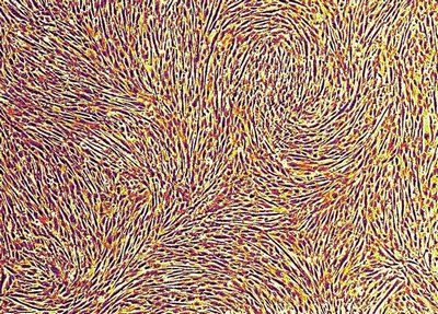
Albert Folch, University of Washington
“Van Gogh’s cells”: A magnified image of muscle cells after about one week of growth, when they start to fuse. The cells have been digitally colored.
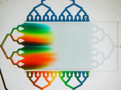
Albert Folch, University of Washington
A microfluidic device is filled by substituting water with dyes, here flowing in from the left. The channels at the top and bottom of the chamber were filled with dyes a minute ago, but are now stopped by two horizontal valves.
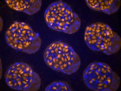
Albert Folch, University of Washington
Researchers prepared this surface using a stretchy rubber stencil with circular holes and filling the holes with a black protein that doesn’t stick to cells. Then they put another stencil and filled it with blue proteins that do stick to cells. But because the stencil was flexible it didn’t quite match up, resulting in the slightly offset circles. Experimenters then attached cells to the sticky blue protein circles left by the stencil. This technique creates surfaces that act like miniature Petri dishes.
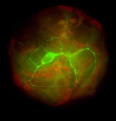
Albert Folch, University of Washington
A neuron from the hippocampus, a section of the brain known to play an important role in the formation of memory. This neuron (green) sits on top of a glial cell (red), a type of brain cell that provides physical support and nutrients to neurons. They are combined in order to understand how glial cells and neurons communicate with each other by looking at just a single cell of each type.
Albert Folch, University of Washington
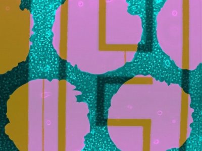
Liver cells (blue-green) grown on top of a gold electrical circuit (yellow) on a sheet of glass (pink). This circuit is a prototype for a device that could measure electrical activity in individual cells.
Albert Folch, University of Washington
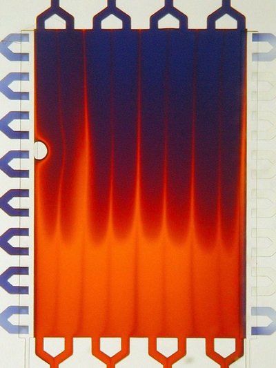
Snapshot of a microfluidic chamber as orange dye is replaced with blue dye. Fluid flows from top to bottom. The small, clear circle is an air bubble. When the researchers see this bubble, they stop the experiment and force the air bubble out. But it makes for a pretty picture.
Albert Folch, University of Washington
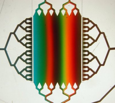
Fluids shoot through tiny valves at the top and bottom to create a rectangular area where the concentration of the fluid changes gradually. The colors help experimenters see how much the fluids are mixing. Flow goes from bottom to top.
Albert Folch, University of Washington
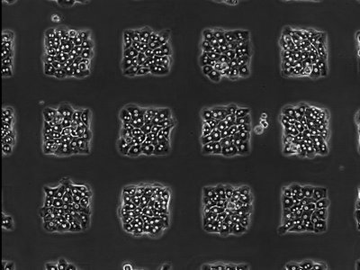
Liver cells imprisoned in square islands. The islands were made by placing a rubber stencil containing square holes on a glass surface and allowing collagen to fill in the holes. Cells only stick to the square-shaped collagen areas.
Albert Folch, University of Washington
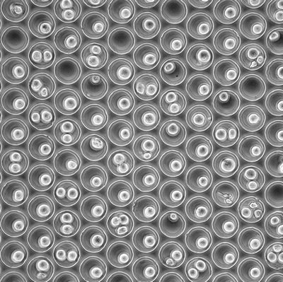
Cells confined to tiny wells. The size of the wells was optimized so that it would fit only one cell. However, since the size of cells varies, you can see that some wells end up holding two or even three cells, while others remain empty.
Albert Folch, University of Washington
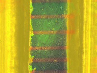
Cells attached to the inside of a channel made of mylar, a type of polyester. The mylar is bright yellow because it becomes fluorescent under light. The red lines are proteins that stick to cells, while the green area is a protein that doesn’t stick to cells. This experiment failed—instead of sticking to the red areas, the cells attached everywhere. Also, the mylar’s fluorescence was an undesired effect.
Albert Folch, University of Washington
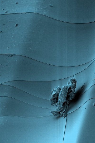
An image of a speck of dust on a piece of rubber, taken with a scanning electron microscope. The rubber was molded to form a half-pipe trench and coated with a delicate layer of metal that cracked accidentally, creating the thin wavy lines.
Albert Folch, University of Washington
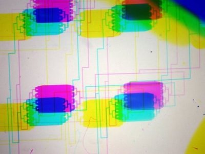
The researcher was shaking the plate while this picture was taken. The camera takes three pictures, each using a different color filter. Because the picture moved the image is repeated in multicolor.
Albert Folch, University of Washington
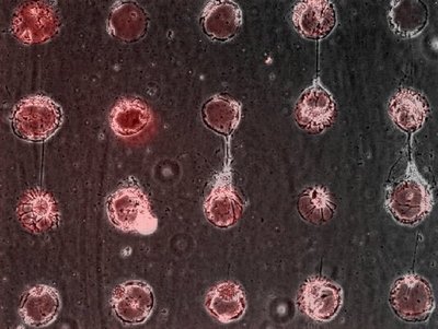
The grid of red dots is made of a fluorescent protein that sticks to neurons. Once attached to a spot, the neurons send axons to nearby islands, forming the same type of synaptic connection that our bodies use to transmit brain and muscle signals.
Albert Folch, University of Washington
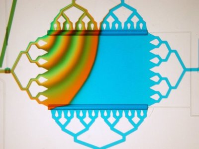
Green, yellow and orange dyes flow from left to top. The blue dye flows up from the bottom. The different currents create a gradient in the central rectangle.
Albert Folch, University of Washington
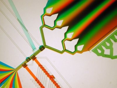
The rainbow in the lower left corner merges into a single channel but the rainbow stays intact. This feeds into a chamber and creates a gradient.
Albert Folch, University of Washington
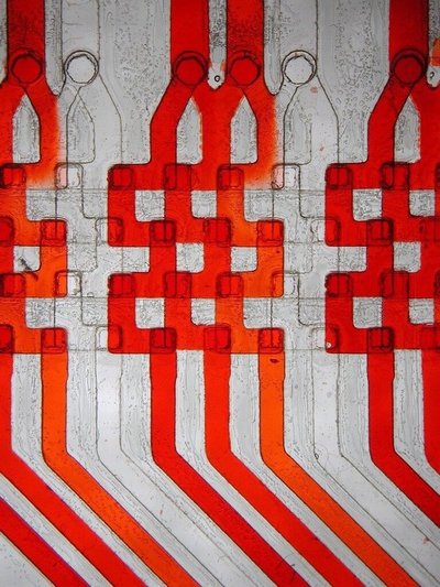
Closeup of a micro-mixing machine. The fluids flow down through four channels that are placed one in front of the other.
Albert Folch, University of Washington
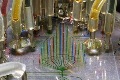
The tubes feed different colored liquids into the microfluidic device. The bottom channel (green) feeds into a larger chamber underneath (not shown). A system of tiny valves lets the researcher choose which liquids to send into the chamber.
Albert Folch, University of Washington
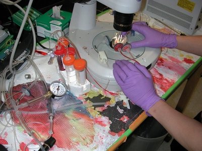
Scientists making a mess! This researcher runs colored liquids through microfluidic devices to see how the liquids are mixing.
Albert Folch, University of Washington
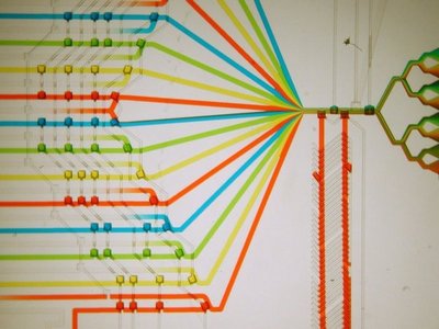
The bumps on the left side are tiny valves. Researchers open the valves to select different combinations of fluid (here, all are opened). Fluids then flow from left to right through this “microfluidic multiplexer.” The fluids mix in the single channel and then flow into the fingerlike channels on the right, which supply a large mixing chamber (not shown).
Albert Folch, University of Washington
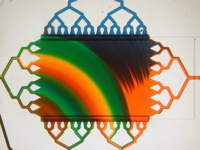
A microfluidic gradient generator in the middle of switching from orange dye to blue dye. Flow enters the chamber from the left and top, and exits through the right and bottom.
