November 24, 2010
“Finger trap” tension stabilizes cells’ chromosome-separating machinery
Scientists have discovered an amazingly simple way that cells stabilize their machinery for forcing apart chromosomes. Their findings are reported Nov. 25 in Nature.
When a cell gets ready to split into new cells, this stable set-up permits its genetic material to be separated and distributed accurately. Otherwise, problem cells — like cancer cells— arise.
The human body contains more than a trillion cells, and every single cell needs to have the exact same set of chromosomes. Mistakes in moving chromosomes during cell division can lead to babies being born with genetic conditions like Down syndrome, where cells have an extra copy of chromosome 21.
“A striking hallmark of cancer cells,” said one of the senior authors of the study, Sue Biggins, an investigator in the Basic Science Division, Fred Hutchinson Cancer Research Center in Seattle, “is that they contain the wrong number of chromosomes, so it is essential that that we understand how chromosome separation is controlled. This knowledge would potentially lead to ways to correct defects before they occur, or allow us to try to target cells with the wrong number of chromosomes to prevent them from dividing again.”
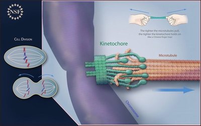
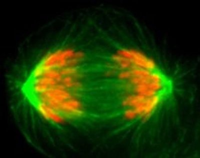
The machine inside cells that moves the chromosomes is the kinetochore. These appear on the chromosomes and attach to dynamic filaments during cell division. Kinetochores drive chromosome movement by keeping a grip on the filaments, which are constantly remodeling. The growth and shortening of the filaments tugs on the kinetochores and chromosomes until they separate.
“The kinetochore is one of the largest cellular machines but had never been isolated before,” Biggins said, “Our labs isolated these machines for the first time. This allowed us to analyze their behavior outside of the cell and find out how they control movement.”
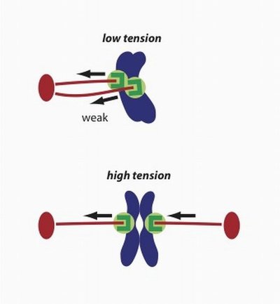
“We demonstrated that attachments between kinetochores and microtubule filaments become more stable when they are placed under tension,” noted Dr. Charles “Chip” Asbury, a University of Washington (UW) associate professor of physiology and biophysics. Originally trained in mechanical engineering, Asbury studies molecular motors in cells. He is also a senior author on the Nov. 25 Nature paper.
Asbury likened the stabilizing tension on the filament to a Chinese finger trap toy — the harder you try to pull away, the stronger your knuckles are gripped.
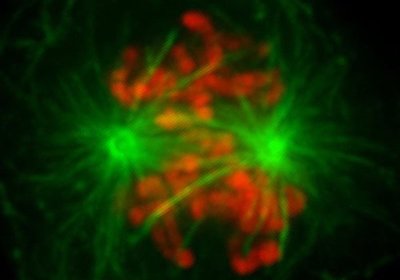
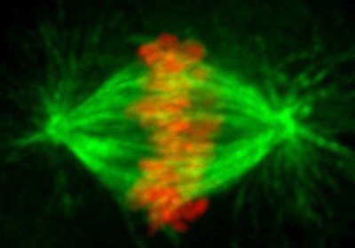
Asbury explained how this tension-dependent stabilization helps chromosomes separate according to plan. As cell division approaches, a mitotic spindle forms, so named by 19th century scientists because the gathering microfilaments resemble a wheel spinning thread.
When chromosome pairs are properly connected to the spindle, with one attached to microtubules on the right and the other to microtubules on the left, the kinetochore comes under mechanical tension and the attachment becomes stabilized, sort of like steadying a load by tightening ropes on either side. This is a simple, primitive mechanism.
“On the other hand,” Asbury said,” if the chromosome pair is not properly attached, the kinetochores do not come under full tension. The attachments are unstable and release quickly, giving another chance for proper connections to form.” Kinetochores are not just connectors, but also are regulatory hubs. They sense and fix errors in attachment. They emit “wait” signals until the microtubule filaments are in the right place.
The research team conducted this study using techniques to manipulate single molecules to see how they worked. These methods allow scientists to take measurements not possible in living cells. The native kinetochore particles were purified from budding yeast cells.
To the best of his knowledge, Asbury said, “Intact, functional kinetochores had not previously been isolated from any organism.” The purification of the kinetochores allowed the research team to make the first direct measurements of coupling strength between individual kinetechore particles and dynamic microtubules.
The results of this study contribute to wider efforts to understand a puzzling phenomenon on which all life depends: How are motion and force produced to move duplicated chromosomes apart before cells divide?
The research was funded by grants from the National Institute of General Science at the National Institutes of Health, the National Science Foundation, the Packard Foundation, the Kinship Foundation and the Beckman Foundation.
In addition to senior authors Biggins and Asbury, the lead authors on this study are Bungo Akiyoshi, from the Molecular and Cellular Biology Program at the UW and the Division of Basic Sciences, Fred Hutchinson Cancer Research Center; and Krishna K, Sarangapani and Andrew F. Powers, both of the UW Department of Physiology & Biophysics. The research team included Christian R. Nelson, Fred Hutchinson Cancer Research Center; Steve L. Reichow, UW Department of Biochemistry; Hugo Arellano-Santoyo, Fred Hutchinson Cancer Research Center, UW Molecular and Cellular Biology Program, and UW Department of Physiology & Biophysics; Tamir Gonen of the UW Department of Biochemistry and the Howard Hughes Medical Institute; and Jeffrey N. Ranish of the Institute for Systems Biology in Seattle.
